Celigo Image Cytometer Training with Revvity @ 953 Indiana St
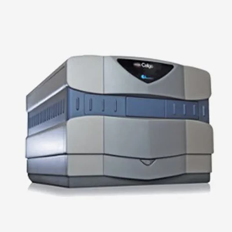
Get hands-on with the Revvity Celigo—a plate-based, benchtop brightfield and fluorescence imaging system for whole-well, live-cell analysis. Celigo images directly in your vessel (6–1536-well plates, T-flasks, 10 cm dishes, and slides) without disturbing cell state, and delivers single-cell-level insights faster than flow and far beyond what ELISA/protein assays can reveal.
Why attend: Leave with a ready-to-use analysis template tailored to your assay (e.g., confluence, colony/spheroid counting, transfection efficiency), so you can hit the ground running on the shared instrument. Limited hands-on seats — registration required.
Who Should Attend
Researchers, scientists, and technicians who plan to use (or are curious about) the Celigo in the MBC BioLabs shared equipment lab. New users welcome.
What You’ll Learn
Whole-well acquisition in plates, flasks, and dishes: focusing, exposure, and channel setup (BF + fluorescence).
Cell-level segmentation & quantification: tuning masks, thresholds, and QC for reliable counts.
Assay workflows: confluence & growth curves, colony/spheroid detection and viability, transfection efficiency, scratch/wound closure.
Batch analysis & reporting: plate maps, template saving, and data export for downstream stats/visualization.
Best practices to maximize speed and reproducibility on the shared instrument.
Agenda
10:00–11:00 AM — Overview & Applications (Cytiva Conference Room, 1st floor)
Brief talk and live walkthrough of Celigo capabilities and common assays.11:00 AM–1:00 PM — Hands-On Training (Core Lab, 953 Indiana St)
Guided imaging on real samples