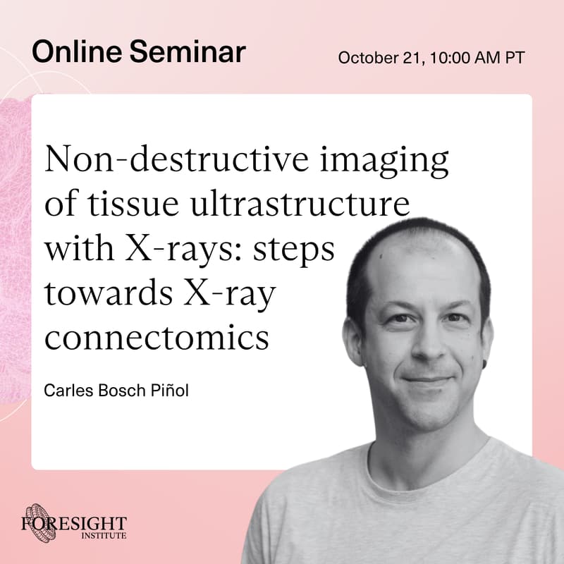

Carles Bosch Piñol | Non-destructive imaging of tissue ultrastructure with X-rays: steps towards X-ray connectomics
Foresight Institute’s Neurotech Group
Non-destructive imaging of tissue ultrastructure with X-rays: steps towards X-ray connectomics
Abstract: Maps of biological tissues at subcellular to ultrastructural detail hold key insights to mechanistically understand how organs and organisms work. In the brain, such 3D maps provide the location, morphology and connectivity of all cells inside - the connectome.
While volume electron microscopy is the current golden standard for connectomics1, hard X-ray nanoimaging brings important advantages:
Hard X-rays can penetrate >mm into heavy metal-stained tissues with preserved ultrastructure, enabling non-destructive 3D imaging without slicing the sample
Phase contrast imaging has resolved 4nm detail in computer chip samples2 and 40nm detail in brain tissue3
4th generation synchrotrons create X-ray beams with enough flux to image petavoxels in days4
Multiple synchrotrons are building tissue nanoimaging beamlines for their 4th generation sources, starting by the European Synchrotron (Grenoble, France) and ALBA (Cerdanyola del Vallès, Barcelona, Spain). A customised design for connectomics enables tailored compromises so the target datasets can be obtained robustly.
In this talk Carles will review how X-ray nanoimaging can be most useful in combination with the rich toolbox that is available to connectomics researchers, highlighting critical adjacent technologies and use cases.
Bio: Carles is a neuroscientist interested in biological tissues. He has specialised in correlative multimodal imaging of neuronal circuits. By combining an array of imaging techniques, he has developed a strategy to explore ultrastructure at targeted regions in the brain while keeping the big picture that provides context.
After earning a degree in biotechnology, he received my PhD at the University of Barcelona for studying how synaptic plasticity is regulated in the adult mouse brain at the transcriptomic, molecular, and structural levels. His research at the Sensory Circuits and Neurotechnology lab at the Francis Crick Institute (London) addresses how the mammalian brain detects odours, by integrating insights on the structure and function of the sensory neuronal circuits underlying perception. He currently leads the design of a nanoimaging and X-ray fluorescence beamline at the ALBA Synchrotron.
Additionally, He is driven to facilitating scientific discovery through multidisciplinary collaboration.
A group of neuroscience researchers, entrepreneurs, and allies advancing beneficial short-term and long-term neurotechnology applications.
Subscribe to our newsletter
Nominate a seminar presenter/topic
Share this application with colleagues who’d like to join
Zoom Link: https://us02web.zoom.us/j/87048895463